Brain organoids derived from human fetal brain tissue provide new tools for studying brain development and disease
Publication Date:2024-01-26
bioGenous Science Focus
This study demonstrates that healthy human fetal brains self-organize into organoids in vitro and can be cultured long-term. FeBO growth requires maintaining tissue integrity, thereby generating an extracellular matrix (ECM) environment similar to that of tissue, ultimately enabling FeBO expansion. This approach reveals that FeBO lineages derived from distinct central nervous system (CNS) regions retain their brain-specific characteristics, enabling exploration of region-specific brain functions. Furthermore, CRISPR-Cas9 technology was employed to generate FeBO lineages with homozygous mutations for brain cancer research. This study demonstrates that FeBOs constitute a complementary CNS organoid platform.
Human organoid structures are three-dimensional (3D) constructs derived from stem cells that mimic the characteristics of their corresponding tissues, including cellular composition, architecture, and function. Previous approaches primarily focused on establishing organoid models from pluripotent stem cells (PSCs) or tissue-specific stem cells (TSCs). Each model type offers distinct advantages. The human brain and its development exhibit unique characteristics, including more complex layered organization and increased cellular diversity. Consequently, brain organoids have thus far been established exclusively from pluripotent stem cells. This process involves attempting to mimic the brain's natural developmental trajectory using specific molecular combinations. Each small-molecule formulation requires extensive research effort, and a long-term expandable 3D brain in vitro model remains unachieved.
In January 2024, Hans Clevers, Benedetta Artegiani, and colleagues published a research paper titled “Human fetal brain self-organizes into long-term expanding organoids” in the journal Cell.
This study directly developed in vitro self-organizing brain organoids—FeBOs (Fetal brain in vitro self-organizes into organoids, FeBOs)—from fetal brain tissue. These laboratory-cultured organoids establish a novel model for studying brain development and provide valuable new tools for investigating brain development-related disorders, including the progression and treatment of brain tumors.
The study demonstrates that healthy human fetal brain tissue self-organizes into organoids in vitro and can be cultured long-term. FeBOs' growth requires maintaining tissue integrity, which generates a tissue-like extracellular matrix (ECM) environment, ultimately enabling FeBOs expansion. This approach reveals that FeBO lineages derived from distinct CNS regions retain their brain-specific characteristics, enabling exploration of location-specific aspects of brain function. Furthermore, CRISPR-Cas9 technology was employed to generate FeBO lineages with homozygous mutations for brain cancer research. This work demonstrates that FeBOs constitute a complementary CNS organoid platform.
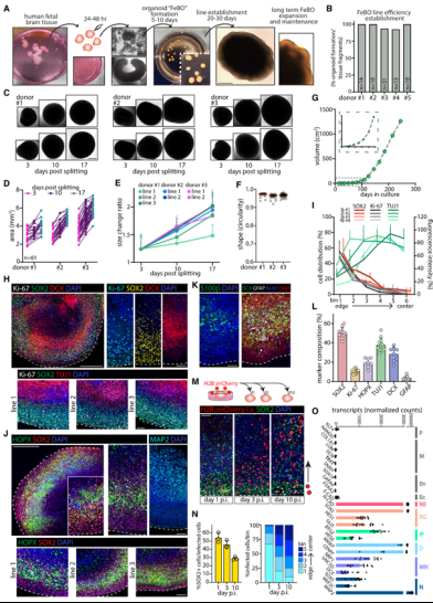
First, the research team successfully established organoid models derived from healthy human fetal brain tissue. Researchers obtained healthy human fetal brain tissue, from which isolated human neural stem cells (NSCs) could grow in culture medium either as a monolayer or as relatively small, disorganized 3D cell aggregates. They then explored whether different culture strategies could produce organoids with tissue-like cellular and structural complexity for long-term expansion. To this end, they dissected the brain tissue into small pieces (1–4 mm in diameter) and cultured them in serum-free, extracellular matrix-free medium. Within the initial 4–8 days, multiple organized 3D structures with distinct boundaries formed. Over time, these structures grew while maintaining an organ-like appearance, termed embryo-derived brain organoids (FeBOs). They found that the success rate for establishing FeBO lines exceeded 95%. By dissecting entire organoid bodies, FeBOs could be reliably passaged, with each dissected fragment capable of regenerating complete organoid bodies. This yielded stably expanded FeBO lines, enhancing reproducibility for downstream applications. When placed in maturation medium, FeBOs slowed their growth, while characteristics associated with later developmental stages, such as glial differentiation, became more pronounced.
Next, the researchers assessed FeBOs' cellular composition using a panel of common neurodevelopmental markers. Results revealed abundant neural stem/progenitor cells (SOX2+) at FeBOs' periphery, while neuronal cells (TUJ1+, DCX+, NeuN+) resided centrally. Quantitative analysis of cell composition revealed regenerative abundance across distinct cell populations, highlighting cellular heterogeneity within FeBOs. They further assessed cell lineage dynamics via lentiviral infection of intact FeBOs, finding that labeled cell numbers increased over time and were also located within neuron-rich regions of the organoid, supporting ongoing neurogenesis. Transcriptomic profiling also demonstrated their unique neural ectodermal characteristics.
Figure 1Establishment of Human Fetal Brain Self-Organizing Organoids
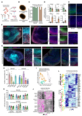
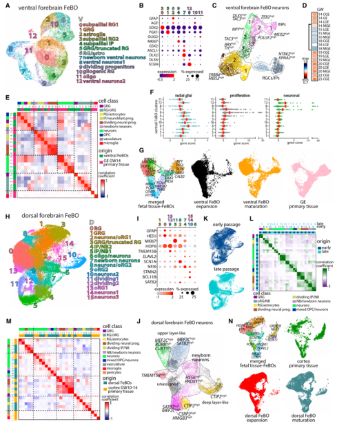
Subsequently, to investigate whether brain region-specific FeBOs could be cultured and whether they retained intrinsic tissue characteristics in vitro, Researchers cultured fragments from distinct brain regions and identified them via qPCR. They found that brain organoids derived from fetal brain tissue could sustain multiple characteristics specific to their brain regions over extended periods. These organoids responded to signaling molecules crucial for brain development. For instance, FeBOs from dorsal and ventral forebrain regions exhibited distinct behaviors when exposed to dorsal signaling molecules, suggesting they may also capture distinct regional responses.
Figure 2 FeBOs retain brain characteristics of the original tissue region
To investigate the cellular heterogeneity of FeBOs in greater detail, researchers performed single-cell sequencing on cells derived from FeBOs. Through subpopulation clustering analysis, they defined the heterogeneity of neurogenic and neuronal populations, identifying distinct types of intermediate neuronal precursors. Simultaneously, they found that FeBOs cells integrated well with human fetal brain tissue cells, suggesting similarities between the two. Furthermore, they compared the similarity between early-stage (2-month) and late-stage (6-month) FeBOs, finding that cells integrated well in the same dimensionality reduction space. Correlation analysis showed high relatedness among most cell subpopulations. These features highlight the cellular heterogeneity exhibited by FeBOs and their high similarity to primary cell types.
Figure 3 Cellular similarity between FeBOs and human fetal brain tissue
Increasing evidence indicates that the extracellular matrix (ECM) and ECM-cell interactions play a crucial role in regulating human brain development. At the transcriptomic level, researchers observed high similarity between FeBOs and human brain tissue development. Further comparison with proteomic data revealed good similarity in transcriptomic-proteomic expression for key ECM brain components such as proteoglycans, glucans, and laminarins. They also investigated functional ECM secretion through mass spectrometry analysis of FeBO supernatant, detecting various secreted ECM components—including those specifically enriched in human fetal brain tissue.
To determine whether FeBOs' intrinsic capacity to secrete tissue-like matrix relates to maintaining intercellular organization and integrity, researchers compared the secretome of intact FeBOs, neurospheres, and FeBOs cultured for 4 days. Results indicate that sustained maintenance of tissue integrity promotes ECM niche formation, which in turn may facilitate the continued expansion of FeBOs within tissue-like structures. Notably, matrix secretion significantly increases shortly after FeBOs division, highlighting the ECM's importance in FeBOs regeneration.
Figure 4Tissue-like ECM niche of FeBOs
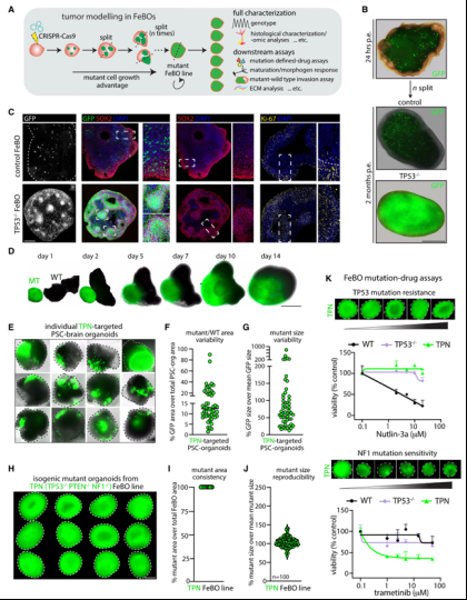
Given the rapid expansion capacity of tissue-derived organoids, the research team further investigated their potential for simulating brain cancer. Using CRISPR-Cas9 gene editing technology, they introduced TP53 gene defects into a small number of cells within FeBOs. Results showed that after three months, TP53-deficient cells completely outnumbered healthy cells within the organoids, indicating that this FeBOs mutant strain acquired the abnormal proliferation characteristics of cancer cells. Simultaneously, CRISPR-Cas9 was employed to inactivate three genes associated with glioblastoma, a type of brain tumor: TP53, PTEN, and NF1. The response of these mutated FeBOs strains to existing cancer drugs was then observed. These mutated FeBOs strains offer the advantages of scalability and reproducibility, enabling large-scale functional screening and providing new tools for investigating the mechanisms of brain tumors and developing treatments.
Figure 5 CRISPR-edited FeBOs can be used for brain tumor modeling
In summary, in this study, the research team demonstrated that small fragments of human fetal brain tissue can be expanded into organoids (FeBOs) under defined culture conditions over extended periods, exhibiting reliable molecular profiles and cellular heterogeneity. Within these FeBOs, stem/progenitor cells localize at the organoid periphery, while neurogenesis occurs centrally, creating a self-sustaining organoid system. This provides a valuable tool for studying brain development and the progression and treatment of brain development-related diseases.