VPS35 Promotes Cell Proliferation via EGFR Recycling and Enhances Gastric Cancer Response to EGFR Inhibitors
Publication Date:2024-01-19
bioGenous Science Focus
The team's research reveals a novel perspective on VPS35's role in activating the ERK/AKT pathway and promoting tumorigenesis in gastric cancer. Mechanistically, this study demonstrates that VPS35 promotes EGFR recycling to the cell surface, potentially amplifying intracellular protein trafficking and receptor transport networks. As VPS35 increases gastric cancer sensitivity to EGFR inhibitors, it may serve as a potential therapeutic biomarker for guiding anti-EGFR therapy in gastric cancer patients.
In February 2023, Bingya Liu's team published an article titled “VPS35 Promotes Cell Proliferation via EGFR Recycling and Enhances EGFR Inhibitor Response in Gastric Cancer” in the journal eBioMedicine. The team's work revealed that VPS35 promotes the initiation and progression of gastric cancer by recycling EGFR to the cell surface and activating downstream pathways. By increasing the density of EGFR on the cell surface, VPS35 enhances the sensitivity of gastric cancer to EGFR inhibitors. These findings suggest that anti-EGFR therapy may be applicable for gastric cancer patients, with VPS35 serving as a potential therapeutic efficacy biomarker.
Gastric cancer ranks fifth in cancer incidence and fourth among the world's leading causes of death. In the team's prior research, a gastric cancer prognosis model was constructed using a functional genomic database generated through CRISPR-Cas9 screening. Among the model's components, the vacuolar protein sorting-associated protein 35 (VPS35) drew particular attention. The reverse complex comprises three major components: VPS35, VPS26, and VPS29. VPS35, which transports proteins from endosomes to the trans-Golgi network, plays a dominant role in cargo selection within the reverse complex. Previous studies indicated VPS35 is a prognostic-associated gene in gastric cancer. However, the carcinogenic mechanism underlying protein transport via VPS35 remains unclear.
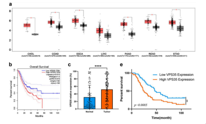
To further investigate VPS35's role in cancer, the team analyzed its expression levels in tumors and adjacent non-tumor tissues across digestive system cancers using GEPIA based on the TCGA database. They found that high VPS35 expression in gastric, colorectal, and pancreatic cancer tumors was positively correlated with shorter patient survival. Higher VPS35 staining scores correlated with deeper local invasion, regional lymph node metastasis, and advanced TNM staging. This suggests that upregulation of VPS35 in gastric cancer tumor tissues is associated with poor prognosis. (Figure 1)
Figure 1VPS35 Expression Levels in Six Digestive System Cancers and Overall Survival Curves for 384 Patients in the Gastric Cancer Cohort
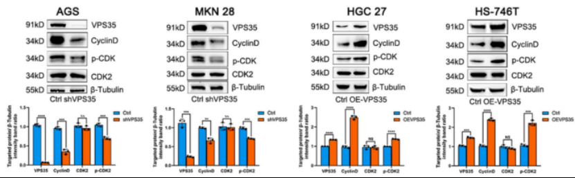
To investigate the effect of VPS35 on gastric cancer cell proliferation, the team established shRNA-transduced cell lines. Compared with the negative control group, the VPS35 low-expression group (AGS, MKN28) exhibited significantly suppressed cell proliferation, whereas overexpression (HGC27, HS-746T) promoted cell proliferation. EdU staining further revealed that VPS35-overexpressing cells exhibited higher EdU positivity compared to VPS35-underexpressing cells. Furthermore, cell cycle analysis revealed that VPS35 accelerates the cell cycle progression and promotes proliferation in gastric cancer cells by activating the CDK2 and cyclin D pathway. The in vivo effects of VPS35 were further investigated using xenograft tumor models, where tumors in the VPS35 low-expression group exhibited lower weights than the negative control group, while those in the high-expression group showed higher weights. (Figure 2)
Figure 2 Expression levels of the indicator protein in VPS35 high- and low-expression group cells
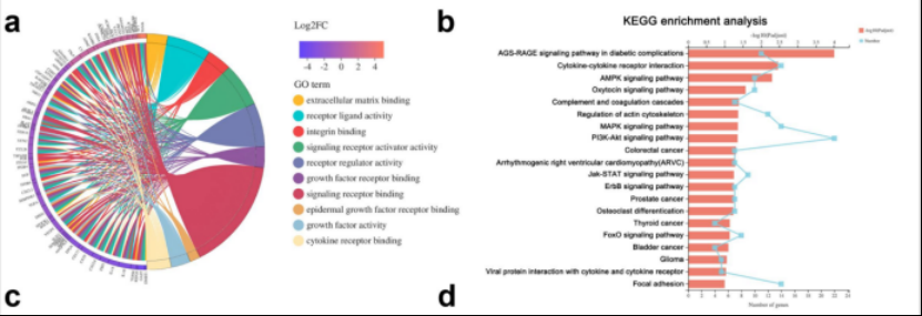
To determine which proteins VPS35 interacts with and the mechanism underlying its proliferation-promoting function, transcriptome profiling was performed in cells with low VPS35 expression and their negative control counterparts. Gene Ontology (GO) enrichment analysis of differentially expressed genes (DEGs) revealed enrichment in receptor-ligand interactions, EGFR-binding-related genes, and epithelial cell proliferation regulation genes. Gene set enrichment analysis indicated that VPS35 expression positively correlates with growth factor receptor binding. (Figure 3)
Figure 3Pathway enrichment analysis of significantly differentially expressed genes between VPS35 low-expression group cells
Subsequently, the team conducted correlation analyses in the TCGA database between VPS35 expression levels and the expression levels of multiple genes related to MAPK-associated receptors. Flow cytometry revealed that only the density of EGFR on the cell surface varied with changes in VPS35 expression. IHC staining confirmed that VPS35 expression levels were positively correlated with EGFR expression in the validation cohort. Co-immunoprecipitation (Co-IP) assays further confirmed the binding interaction between VPS35 and EGFR. These findings indicate that VPS35 binds to EGFR, and its overexpression increases surface EGFR density.
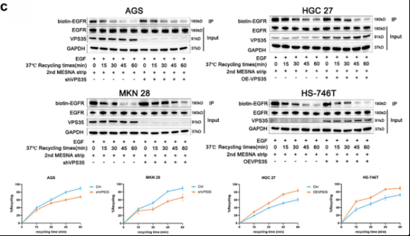
To determine whether VPS35 transports EGFR back to the membrane and inhibits EGFR degradation by binding to it, the team performed EGFR Western blot analysis and assessed EGFR recycling using a cell surface biotinylation assay. They found that VPS35 slows EGFR degradation by promoting its recycling to the cell surface. (Figure 4)
Figure 4Recycling rate of EGFR at different time points for each cell group
The team discovered that increased EGFR signaling, driven by VPS35 upregulation and elevated EGFR levels on the cell surface, is transmitted to the cytoplasm, leading to cell proliferation. Following EGFR knockdown, VPS35 upregulation could no longer promote cancer cell growth. It was concluded that VPS35 activates downstream EGFR pathways, thereby promoting tumor growth.
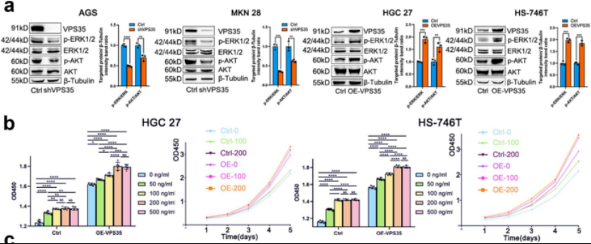
Given that EGFR inhibitors primarily fall into two categories—anti-EGFR monoclonal antibodies (cetuximab) and small-molecule tyrosine kinase inhibitors (erlotinib)—the team also tested erlotinib sensitivity in VPS35-overexpressing cells. Consistent with previous findings, VPS35 overexpression reduced the IC50 value of erlotinib, while low expression increased it. Patient-derived organoids (PDOs) further confirmed that VPS35 enhances gastric cancer sensitivity to erlotinib. Tumors from Patient 2 (P2) exhibited low VPS35 expression, whereas those from Patient 5 (P5) showed high VPS35 expression. P2 organoids contained fewer Ki67-positive cells than P5 organoids. These findings indicate that VPS35 overexpression sensitizes gastric cancer cell lines and organoids to erlotinib, suggesting VPS35 levels may guide clinical application of erlotinib. Furthermore, erlotinib significantly reduced tumor growth in the high-VPS35 PDX#2 group, whereas tumor growth in the low-VPS35 PDX#1 group showed no inhibition. Erlotinib-treated PDX#2 tumors were smaller than vector tumors. These findings indicate that VPS35 plays an oncogenic role in preclinical models of gastric cancer and enhances sensitivity to erlotinib. (Figure 5)
Figure 5Phosphorylation levels of ERK1/2 and AKT in each group and CCK8 assay results
The team's research reveals a novel perspective on VPS35's role in activating the ERK/AKT pathway and promoting tumorigenesis in gastric cancer. Mechanistically, this study demonstrates that VPS35 promotes the recycling of EGFR to the cell surface, potentially amplifying intracellular protein trafficking and receptor transport networks. As VPS35 increases gastric cancer sensitivity to EGFR inhibitors, it may serve as a potential therapeutic biomarker for guiding anti-EGFR therapy in gastric cancer patients.